Tumorigenesis and regeneration in Drosophila
Research summary:
Using Drosophila as a model system our work has focussed primarily on the study of two major processes, regeneration and tumorigenesis, and in particular the role of the Jun N-terminal Kinase (JNK) pathway, a conserved pathway known to be associated with tumorigenesis and regeneration in metazoans.
The JNK signalling pathway is a principal inducer of apoptosis in Drosophila, an autocrine function aimed to eliminate damaged or aberrant cells. This pro-apoptotic function includes the elimination by cell competition of oncogenic cells that may appear in development. Recent work has shown that JNK also has a pro-proliferation function, mediated by paracrine signalling and associated with the activity of the JAK/STAT, Wingless (Wg) and Decapentaplegic (Dpp).
Regarding the role of JNK function in tumorigenesis, we have found that it is a major factor in the process. In previous publications (Ballesteros-Arias et al 2014) we proposed that JNK-mediated proliferative signals are responsible for the growth of tumours generated by oncogenic mutations. More recent work (Pinal et al 2018) has demonstrated the full tumorigenic potential of JNK: in absence of apoptosis a transient stress (e.g. ionising radiation) results in persistent JNK activity and of its target pathways JAK/Stat, Wg or Dpp. The end result is continuous paracrine proliferative signalling, which causes tumour overgrowths. Moreover, we have found that the persistent JNK activity after irradiation is due to a maintenance loop involving Reactive Oxygen Species (ROS) and the DUOX maturation factor Moladiev (Mol). It is the existence of this loop what makes the absence of apoptosis potentially tumorigenic. We are in the process of studying JNK gene targets responsible for the tumorigenic response; a recent RNAseq screen performed in the lab has revealed a number JNK targets, several of which are conserved and also associated with human cancer.
We are also investigating the role of JNK in the tumorigenic processes generated by mutations like scribble, erupted or polyhomeotic. These mutations alter respectively apico-basal polarity, endocytosis or identity of epithelial cells and are also conserved in humans. The mechanisms by which defects in such different functions cause tumorigenesis are still obscure.
We are also trying to elucidate the mechanism of JNK activation during cell competition, which is still unclear. Some of the upstream factors are known (flower, sas/ptpd10, spz/toll, azot) but their specific molecular roles are not clear. It is also possible that there may be different ways to activate JNK.
JNK signalling displays also a paracrine function in regeneration processes. We propose to study the regenerative potential of imaginal discs or specific regions of the discs in relation to the local activity of JNK. Regarding regeneration we have compared the regeneration potential of the trunk region of the disc (the thorax) with that of the appendage (the wing). A principal finding is that the wing region shows a strong regeneration capacity while the trunk area does not. Our results indicate that this is due to local differences in JNK function, which appears to activate different target genes in trunk and appendage. This finding may be of interest to solve a classical problem in the regeneration field: why some organs are more regenerative than others or why different animal groups exhibit distinct regeneration potential. Furthermore, we have provided the first direct demonstration that the proliferative response required for regeneration is mediated by JNK activity emanating from damaged cells. An additional point of interest is that the trunk and the appendage of the wing disc are developmentally isolated in spite of sharing lineage: they cannot be reprogrammed to generate each other. These results have been published recently (Martin et al 2017; Martin and Morata 2018).
Another goal in the laboratory is to study chromatin remodelling during tissue regeneration. Surviving cells of damaged tissues need to readapt their status for new tissue formation. They do so by remodelling their chromatin to activate or repress a new battery of genes. A recent report based on work with mouse embryos suggest that activation of retrotransposon LINE elements regulates global chromatin accessibility at the beginning of development (Jachowicz, et al., 2018) suggesting that retrotransposon activation is integral to the early developmental program. During regeneration, nuclear chromatin might acquire an “intermediate” state that resembles the young embryonic totipotent state and that is mediated by retrotransposons.
Previous results in the laboratory show that the Roo and F-Element retrotransposons of Drosophila suffer profound changes in their leveles in the first hours of embryonic development. We are in the process of measuring the levels of these retrotransposon during tissue regeneration as well as altering them to study their putative role in regeneration.
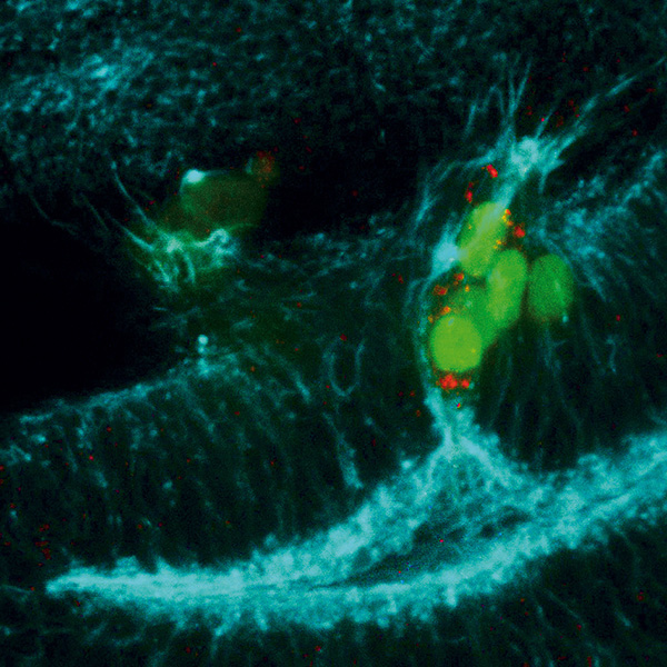
Drosophila wing disc containing a marked clone (green) deficient for the pro-apoptotic genes and over-expressing the JNK pathway. The cells of the clone express the metalloprotease 1, which degrades extracellular matrix. Note the presence of F-actin protrusions (blue) at the basal region of the cells.
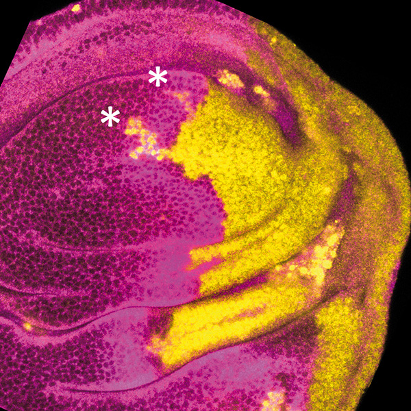
Wing imaginal disc in which posterior compartment cells (right side) have suffered a treatment of 2 days of apoptosis induction followed by 3 days of recovery. The cells belonging to the posterior lineage are labeled in yellow, while the anterior compartment is labeled in magenta. Note the presence of groups of cells (asterisks) that were originated in the posterior compartment and now belong to the anterior compartment. This result suggests that the ablation treatment provokes a transient collapse of the compartments boundary and a change in cell identity.
| Last name | Name | Laboratory | Ext.* | Professional category |
|---|
Relevant publications:
- Javier Menendez; Ainhoa Perez-Garijo; Manuel Calleja; Ginés Morata. 2010. A tumour-suppressing mechanism in Drosophila involving cell competition and the Hippo pathway. PNAS 107 14651-14656.
- Ginés Morata; Evgeny Shlevkov; Ainhoa Perez-Garijo. 2011. Mitogenic signalling from apoptotic cells in Drosophila. Development, Growth and Differentiation 53, 168-176.
- Eveny Shlevkov; Ginés Morata. 2012. A p53-JNK-dependant feedback amplification loop is essential for the apoptotic response in Drosophila. Cell Death & Differentiation 19, 451-460.
- Salvador C Herrera; Raquel Martin; Ginés Morata, G. 2013. Tissue homeostasis in the wing disc of Drosophila: immediate response to massive damage during development. PLoS Genet 9 (4) e10034446.
- Luna Ballesteros-Arias; Veronica Saavedra Ginés Morata. 2014. Cell competition may function either as tumour-suppressing or as tumour-stimulating factor in Drosophila. Oncogene 33, 4377-4384.
- Salvador C Herrera; Ginés Morata G. 2014. Transgressions of compartment boundaries and cell reprogramming during regeneration of the imaginal discs of Drosophila. eLife. 01831.
- Cantero, W., Zaballos, M.A. N. Azpiazu (2015). The Hox/TALE transcription factor Homothorax functions to assemble constitutive heterochromatin during Drosophila embryogenesis. PLoS ONE 10(3):e0120662. doi:10.1371/journal.pone.0120662
- Ginés Morata; Salvador C Herrera. 2016. Cell reprogramming during regeneration in Drosophila: transgression of compartment boundaries. Current Opinion in Genetics & Development 40, 11-16.
- Antonio Montes; Ginés Morata. 2017. Homeostatic response to blocking cell division in Drosophila imaginal discs: role of the Fat/Dachsous (Ft/Ds) pathway. Dev. Biol 424, 113-123.
- Raquel Martin; Noelia Pinal; Ginés Morata, G. 2017. Regeneration potential of the trunk and the appendages of Drosophila meditated by JNK signalling. Development 144, 3946-3956.
- Noelia Pinal; Maria Martin; Izarne Medina; Ginés Morata G. 2018. Short-term activation of the Jun-N terminal Kinase pathway in apoptosis-deficient cells of Drosophila induces tumorigenesis. Nature Communi. 9(1), 1541.
- N. Azpiazu D. Blom-Dah . 2018 “The Pax protein Eyegone (Eyg) interacts with the pi- RNA component Aubergine (Aub) and controls egg chamber development in Drosophila” Dev. Bio 434, 267-277.
- Noelia Pinal; Manuel Calleja; Ginés Morata. 2019. Pro-apoptotic and pro-proliferation functions of the JNK pathway of Drosophila: roles in cell competition, tumourigenesis and regeneration. Open Biol. 9 (3), DOI: 10.1098/rsob.180256
- Manuel Calleja; Ginés Morata. 2019. Cell competition and tumorigenesis in the imaginal discs of Drosophila. Semin. Cancer Biol. DOI:10.1016/j.semcancer.2019.06.010
Doctoral theses:
- Evgeny Shlevklov (2011) Nuevos mecanismos de regulación de la apoptotis en Drosophila melanogaster. Universidad Autónoma de Madrid. Director: Ginés Morata Pérez.
- Javier Ménendez González (2011) Competición celular y desarrollo de tumores en discos imaginales de Drosophila. Universidad Autónoma de Madrid. Director: Ginés Morata Pérez.
- Salvador C. Herrera (2013) Respuesta homeostática e inducción de cambios de identidad tras ablaciones en Drosophila melanogaster. Universidad Autónoma de Madrid. Director: Ginés Morata Pérez.
- Luna Laura Ballesteros Arias (2016)The role of cell competition in tumour initiation and progression in Drosophila melanogaster . Universidad Autónoma de Madrid. Director: Ginés Morata Pérez.
- María Martin Montero (2016) La apoptosis como factor promotor de tumorogenésis en Drosophila melanogaster. Universidad Autónoma de Madrid. Director: Ginés Morata Pérez.
- Antonio Jóse Montes Ruiz (2016) Control del crecimiento en los discos imaginales de Drosophila melanogaster. Universidad Autónoma de Madrid. Director: Ginés Morata Pérez.
Prizes and distinctions:
- Ginés Morata. Prince of Asturias Prize in Science and Technology, 2007/Premio Príncipe de Asturias de Ciencia y Tecnología 2007
- Ginés Morata. Honorary Doctorate by the Universidad de Alcalá, 2007/Doctorado Honoris Causa por la Universidad de Alcalá, 2007
- Ginés Morata. Honorary Doctorate by the Universidad de Almeria, 2008/Doctorado Honoris Causa por la Universidad de Almería, 2008
- Ginés Morata. National Prize of Genetics, 2015/Premio Nacional de Genética 2015
- Ginés Morata. Science Award (shared with P.A. Lawrence) Centenary Awards of the BritishSpanish Society 1916-2016/Premio (compartido con Peter Lawrence) del Centenario de la Sociedad Hispano Británica
- Ginés Morata. Foreign Member of the Royal Society, 2017/Miembro extranjero de la Royal Society
- Ginés Morata. Foreign Member of the US National Academy of Sciences, 2018/ Miembro extranjero de la Academia Nacional de Ciencias de USA