
Cell biology of inflammation
Research summary:
Inflammation is a primary response to infection, stress and injury. Pathological inflammation can lead to chronic diseases, such as atherosclerosis, multiple sclerosis and hepatitis, and to acute diseases, such as sepsis, which can cause organ failure and death. Long-term inflammatory responses are orchestrated by the secretion of soluble mediators that stimulate cells in the surroundings of an inflammatory focus. A major effect of these mediators is disrupting endothelial and epithelial barriers in the tissue parenchyma to facilitate the passage of soluble molecules and immune cells towards inflammatory sites. Our main objective is to investigate the effect of inflammatory mediators on cell barrier function and, reciprocally, to elucidate how endothelial and epithelial barriers regulate the inflammatory response.
Endothelial cells line the inner surface of the vascular wall, where they form a selective barrier that controls the passage of cells and solutes between the blood and the parenchyma. Our group is interested in investigating the effect of TNF, a cytokine that is central to the inflammatory response, on endothelial barrier function. In recent years we have found that, first, TNF increases de expression of RhoB, a member of the RhoA subfamily of GTPases. In an inflammatory context, high levels of RhoB protein debilitates the endothelial barrier by negatively regulating plasma membrane extensions necessary to form cell-cell junctions (Marcos-Ramiro, JCB, 2016). Second, we have also found that human endothelial beds express at least 24 members of the cadherin superfamily of junctional adhesion proteins (Colás-Algora, CMLS, 2019). Amongst them, Vascular endothelial (VE)-cadherin has the highest expression levels (Colás-Algora, CMLS, 2019) (Figure 1) so we are currently investigating the effect of TNF on this cadherin. We have found that TNF increases endothelial permeability and notably reduces VE-cadherin half-life at junctions. Importantly, TNF also activates a compensatory response by increasing de novo synthesis of VE-cadherin through a pathway involving the transcription factors NF-kb and ETS1, thereby altering VE-cadherin turnover, but maintaining its expression levels to prevent endothelial barrier collapse (Colás-Algora, García-Weber, CMLS, 2019). Finally, we are investigating the role of the three RhoA GTPase subfamily members in preserving VE-cadherin stability and endothelial barrier function, which has led us to device molecular tools to reduce organ edema in a preclinical model of sepsis (patent 201930571).
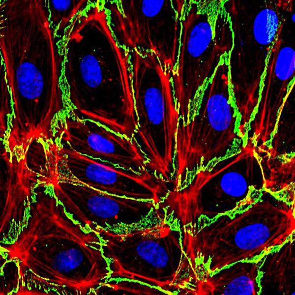
Figure 1. Human endothelial cells from umbilical cords forming a cellular barrier sealed by cell-cell junctions of VE-cadherin (green) bound to filamentous actin (F-actin). Cell nuclei are stained with DAPI (blue).
Once leukocytes extravasate, they establish adhesions with parenchymal epithelial barriers, searching for the inflammatory focus and for dysfunctional cells. The liver parenchyma is composed of irrigated barriers of polarized hepatocytes and cholangiocytes, which constitute the hepatic epithelial cells. Leukocyte infiltration in the hepatic parenchyma is essential for immune-surveillance, control of cancer and infections and tissue regeneration. We are currently studying the role of hepatic epithelial apicobasal polarity in regulating hepatic epithelial interaction with immune cells (Reglero-Real. Cell Rep, 2014). To this aim, we are generating an experimental model of human and murine hepatocyte organoids in 3D derived from liver bipotent stem cells (Figure 2).
Finally, the cornea is one of the most transplanted tissues and the paradigm of tissue organized in cellular barriers. In collaboration with Fundación Jiménez Díaz and the company Cornea Project, we are investigating the organization of corneal endothelial and epithelial barriers and their responses to inflammatory and mechanical stress.
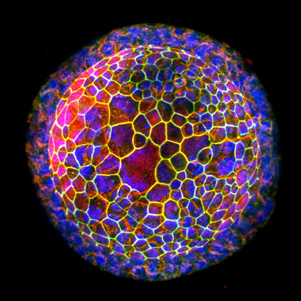
Figure 2. Murine hepatic organoid with an spherical shape, obtained by expanding bipotent hepatic stem cells, cultured in 3D and differentiated to hepatocytes using an specific culture medium, Cells forming this spherical barrier can be distinguished thank to the staining of ZO-1, a component of tight junctions (green), the filamentous actin (F-actin), in red, and nuclei (blue).
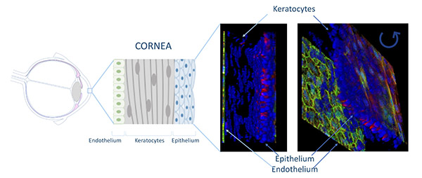
Figure 3. Cornea is one of the most transplanted tissues and a paradigm of cellular barrier organization. Corneas are exposed to multiple inflammatory challenges.
| Last name | Name | Laboratory | Ext.* | Professional category | |
|---|---|---|---|---|---|
| Barroso Fernández | Susana | 324 | 4646 | sbarroso(at)cbm.csic.es | M2 |
| Finis | Domnica | 322 | 4646 | Estudiante TFG | |
| Millán Martínez | Jaime | 324 | 4713 | jmillan(at)cbm.csic.es | E.Científicos Titulares de Organismos Públicos de Investigación |
| Rivas Hidalgo | Gema de | 324 | 4646 | gderivas(at)cbm.csic.es | Técnico Sup. Actividades Tec. y Profes.GP3 |
Relevant publications:
- Cacho-Navas, C., Reglero-Real, N., Colás-Algora N., Susana Barroso, S., de Rivas, G., Stamatakis, K., Feito, J., Andrés G., Fresno M., Kremer, K., Correas I., Alonso M.A., Millán, J*. Plasmolipin regulates basolateral-to-apical transcytosis of ICAM-1 and leukocyte adhesion in polarized hepatic epithelial cells. Cell Mol Life Sci 2022 Jan 9;79(1):61.
- Maeso-Alonso, L. Alonso-Olivares, H., Martínez-García, N., Lopez-Ferreras, L., Villoch-Fernández, J.uente-Santamaría, L., Colas-Algora, N., Fernández-Corona, A., Lorenzo-Marcos, M.E. Jiménez, B., Holmgren, L., Wilhelm, M., Millan, J., del Peso, L., Claesson-Welsh, L., Marques, M.M. and Marin M.C. p73 is required for vessel integrity controlling endothelial junctional dynamics through Angiomotin. Cell Mol Life Sci 2022
- Santaterra, V.A.G, Marx Luz Fiusa, M.M. , Wilfried Hounkpe, B., Chenou F, Vincent Tonasse W., Nilkenes Gomes da Costa, L, Garcia-Weber D, de Farias Domingos, I., de Lima, F., Borba-Junior I, da Silva Araújo, A, Lucena-Araújo A.R. Cavalcante Bezerra M.A., Nunes Dos Santos M.N., Ferreira Costa F, Millán J, De Paula,E.V. Endothelial Barrier Integrity Is Disrupted In Vitro by Heme and by Serum From Sickle Cell Disease Patients. Front Immunol, 2020, 11, 535147
- Colás-Algora N*, García-Weber D*, Cacho-Navas C, Barroso S, Caballero A, Ribas C, Correas I & Millán J. Compensatory Increase of VE-cadherin Expression Through ETS1 Regulates Endothelial Barrier Function in Response to TNFα. Cellular and Molecular Life Sciences. 2019 Aug 8 [Online ahead of print]. DOI: 10.1007/s00018-019-03260-9
- Colás-Algora N & Millán J, How Many Cadherins Do Human Endothelial Cells Express? Cellular and Molecular Life Sciences. 2019. 76 (7), 1299-1317. DOI: 10.1007/s00018-018-2991-9
- García-Weber D and Millán, J. Parallels between single cell migration and barrier formation: the case of RhoB and Rac1 trafficking. Small GTPases. 2018, 9 (4), 332-338. DOI: 10.1080/21541248.2016.1231655
- Marcos-Ramiro B, García-Weber D, Barroso S, Feito, J, Ortega MC, Cernuda-Morollón, E, Reglero-Real N, Fernández-Martín L, Alonso MA, Correas I, Ridley AJ and Millán J. RhoB controls endothelial barrier recovery by inhibiting Rac1 trafficking to the cell border. Journal of Cell Biology; 2016, 213(3):385-402. DOI: 10.1083/jcb.201504038
- Ortega MC, Santander-García D, Marcos-Ramiro, B, Barroso S, Cox S, Jiménez-Alfaro I and Millán J. Activation of Rac1 and RhoA preserve corneal endothelial barrier function. Investigative Ophthalmology & Visual Science 2016, Vol.57, 6210-6222. doi:10.1167/iovs.16-20031
- Santander-García D, Ortega MC, Benito-Martínez S, Barroso S, Jiménez-Alfaro I and Millán J. A human cellular system for analyzing signaling during corneal endothelial barrier dysfunction. Experimental Eye Research. October 2016. http://dx.doi.org/10.1016/j.exer.2016.09.010
- Reglero-Real R, García-Weber D and Millán J. Cellular Barriers after Extravasation: Leukocyte Interactions with Polarized Epithelia in the Inflamed Tissue. Mediators of Inflammation; 2016, http://dx.doi.org/10.1155/2016/7650260.
- Rodríguez-Fraticelli AE, Bagwell J, Bosch-Fortea M, Boncompain G, Reglero-Real N, García-León MJ, Andrés G, Toribio ML, Alonso MA, Millán J, Perez F, Bagnat M, Martín-Belmonte F.Developmental regulation of apical endocytosis controls epithelial patterning in vertebrate tubular organs. Nature Cell Biology, 2015 Mar;17(3):241-50
- Marcos-Ramiro B, García-Weber D, Millán J. TNF-induced endothelial barrier disruption: beyond actin and Rho. Thrombosis and Haemostasis; 2014, 112(6):1088-10.
- Reglero-Real R, Álvarez-Varela A, Cernuda-Morollón E, Feito J, Marcos-Ramiro B, Fernández-Martín L, Gómez-Lechón MJ, Muntané J, Sandoval P, Majano PL, Correas I, Alonso MA and Millán J. Apicobasal polarity controls lymphocyte adhesion to hepatic epithelial cells. Cell Reports; 2014 ;8(6):1879-93.
Doctoral theses:
- Natalia Reglero-Real (JAE) (2013). Función de la polaridad apicobasal de las células hepáticas en la adhesion linfocitaria. Implicaciones en la respuesta inflamatoria del hígado. Sobresaliente Cum Laude.
- Beatriz Marcos-Ramiro (FPI) (2015) (Best thesis award at the CBMSO, academic year 2015-2016). La GTPasa endosomal RhoB regula la recuperación de la barrera endotelial durante la inflamación. Sobresaliente Cum Laude.
- Diego García-Weber (FPI) (2017). Estudio de los mecanismos dependientes e independientes de uniones adherents que regulan la función de barrera endotelial durante la inflamación. Sobresaliente Cum Laude.
- Diana Santander-García, MD (2017). La línea celular B4G12 como modelo para el estudio de la función de barrera del endotelio de córnea. Sobresaliente Cum Laude.
Patents:
- Compuesto para su uso en el tratamiento y/o prevención de septicemia. (patente 201930571 OEPM).