
Scientific facilities
Advanced light microscopy
Facility Head

Álvaro Sahún Español
Scientific Lead

Francisco Javier Díez Guerra
Contact details
91 196 46 43
smoa (at) cbm.csic.es
Booking system
Fees
Reagents
Comments & complaints
Guidelines for Acknowledgments & Authorship
Guidelines for good practices in Material & Method writing
The Advanced Optical Microscopy Facility (SMOA) of the Centro de Biología Molecular Severo Ochoa (CBM) provides advice, technical support and training in advanced optical microscopy and image analysis. Currently, more than 350 users, both from the CBM and other public (research centres, hospitals) and private (biotechnology companies) institutions, benefit from these services. The SMOA provides as well microscopy-related reagents, such as antibodies, probes, dyes, mounting media or imaging chambers.
In addition, SMOA’s technical personnel remains informed about cutting-edge developments in optical microscopy and leads efforts to secure the acquisition of new equipment, ensuring the facility remains at the forefront of technological innovation while meeting the experimental needs of its users. Resulting from this effort, the SMOA offers wide range of state-of-the-art equipment designed to address a variety of optical microscopy techniques:
- Six widefield systems ideal for studies on live and fixed samples, allowing high speed and excellent sensitivity for brightfield, phase contrast, fluorescent and colour (immunohistochemistry) imaging.
- Five point scanning confocal microscopes, which include up to six laser lines and the ability to perform a wide range of photomanipulation experiments and spectral acquisition.
- A spinning disk confocal microscopy system, which maintains confocality at high speeds and features optical photon reasignment module (SoRa) for resolution enhancement.
- A laser depletion-based super-resolution (STED) confocal microscope, together with a fluorescence-lifetime imaging microscopy module (FLIM).
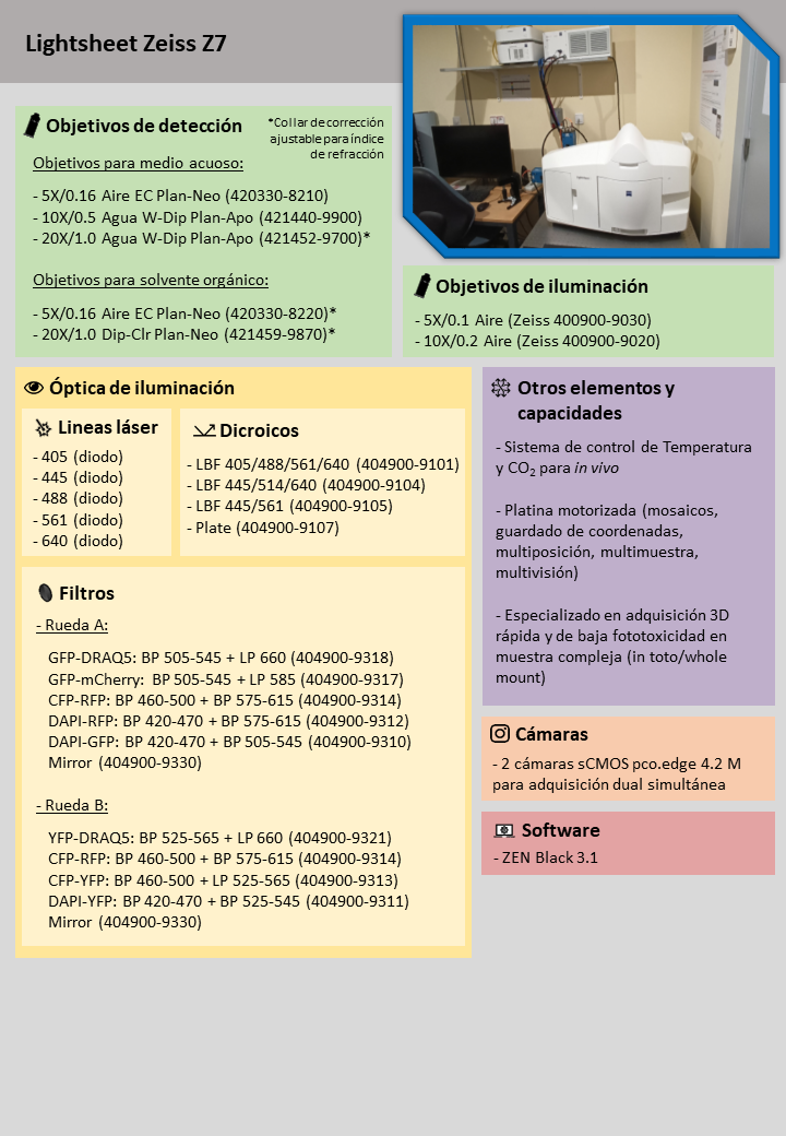
- A lightsheet microscopy system, designed for fast three-dimensional acquisitions with minimal phototoxicity.
Additionally, the SMOA is equipped with a vibratome for sample sectioning, a microfluidic and controlled perfusion system, a 3D printer, and three stereomicroscopes: two with white light and digital camera adapters (ideal for sample preparation and handling), and one with fluorescent light for routine observations.
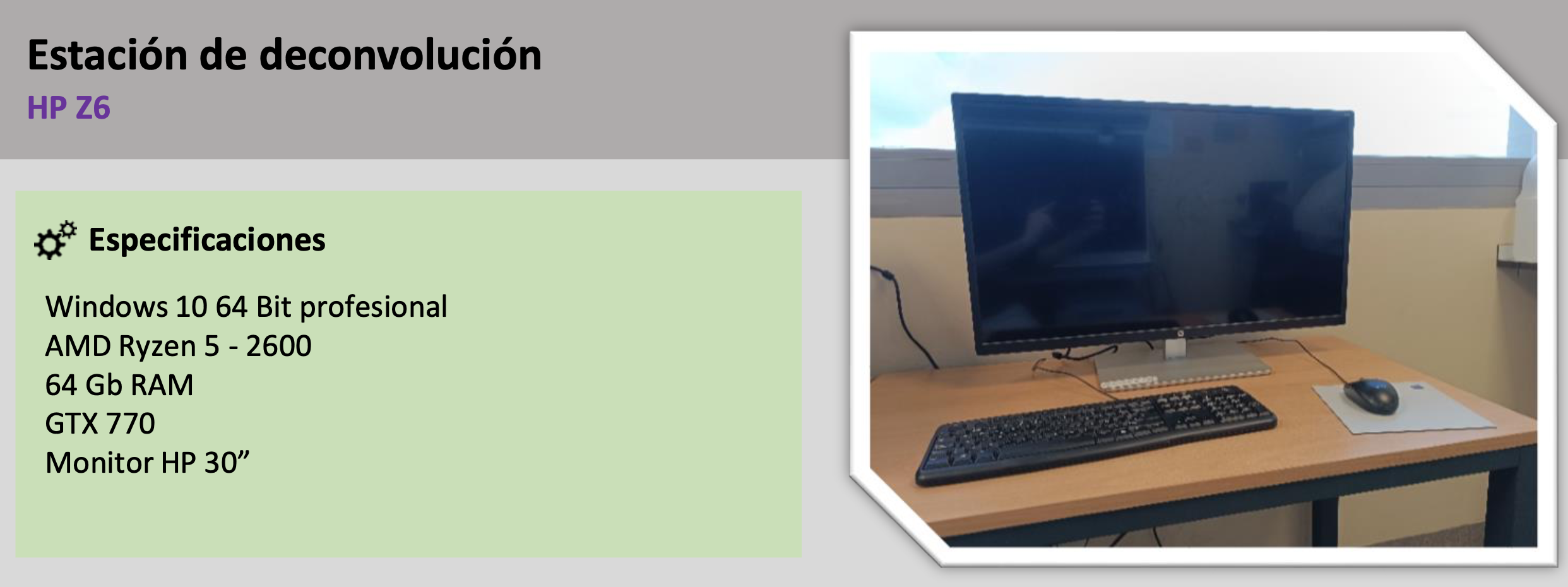
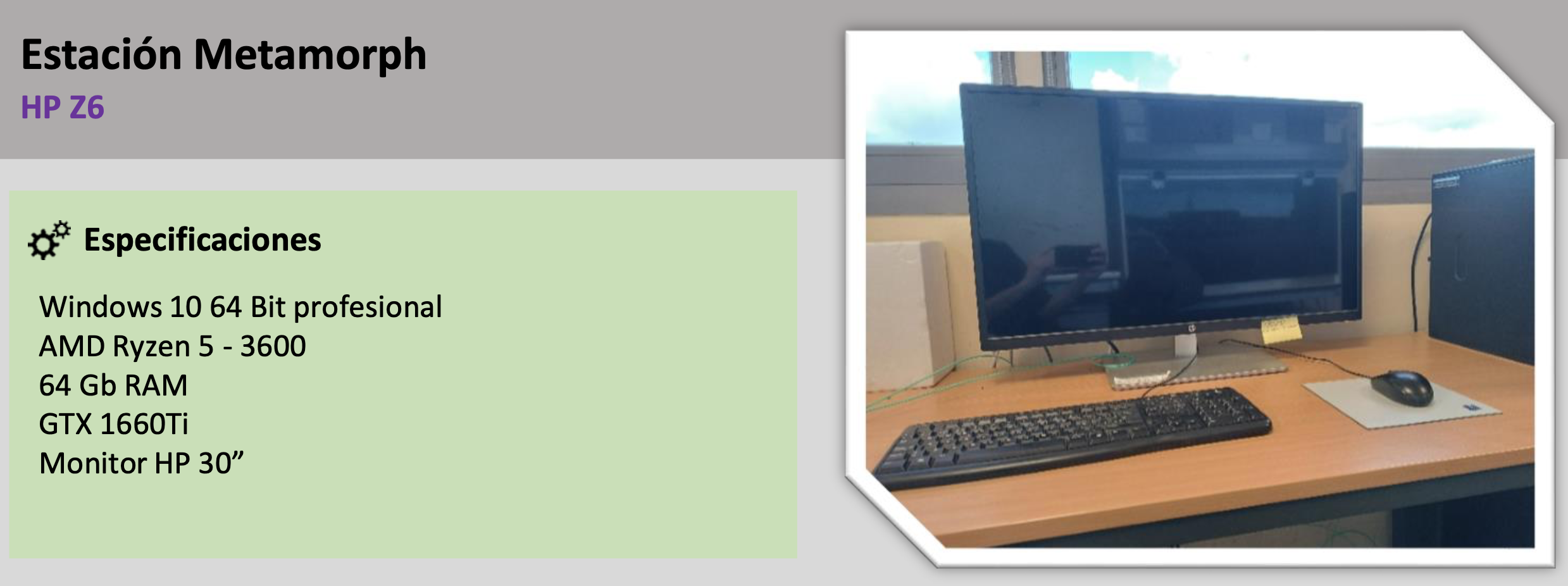
Finally, the SMOA manages two high-performance computer stations, equipped with a wide range of specialised software (ImageJ/Fiji, Huygens, Arivis, CellSense) for data visualisation and analysis.
The SMOA is part of the Madrid Advanced Microscopy Center (MAdMiC) EuroBioImaging Node alongside the Preclinical Biomedicine Facility at the Centro de Biología Molecular Severo Ochoa (CBM) and the Advanced Optical Microscopy and the Cryo-Electron Microscopy Facilities at the Centro Nacional de Biotecnología (CNB).
The SMOA also belongs to the Network of Laboratories and Infrastructures (RedLab) of the Community of Madrid (RLAB-004) and is integrated in the Spanish Network of Advanced Optical Microscopy (REMOA). Since 2009, it holds an ISO9001:2015 quality certification issued by AENOR.
To access the SMOA systems or to purchase reagents and materials related to microscopy, it is necessary to register in our online management system. To create an account, please contact the SMOA staff through our email or phone.
Your Title Goes Here
Your content goes here. Edit or remove this text inline or in the module Content settings. You can also style every aspect of this content in the module Design settings and even apply custom CSS to this text in the module Advanced settings.
Widefield microscopy
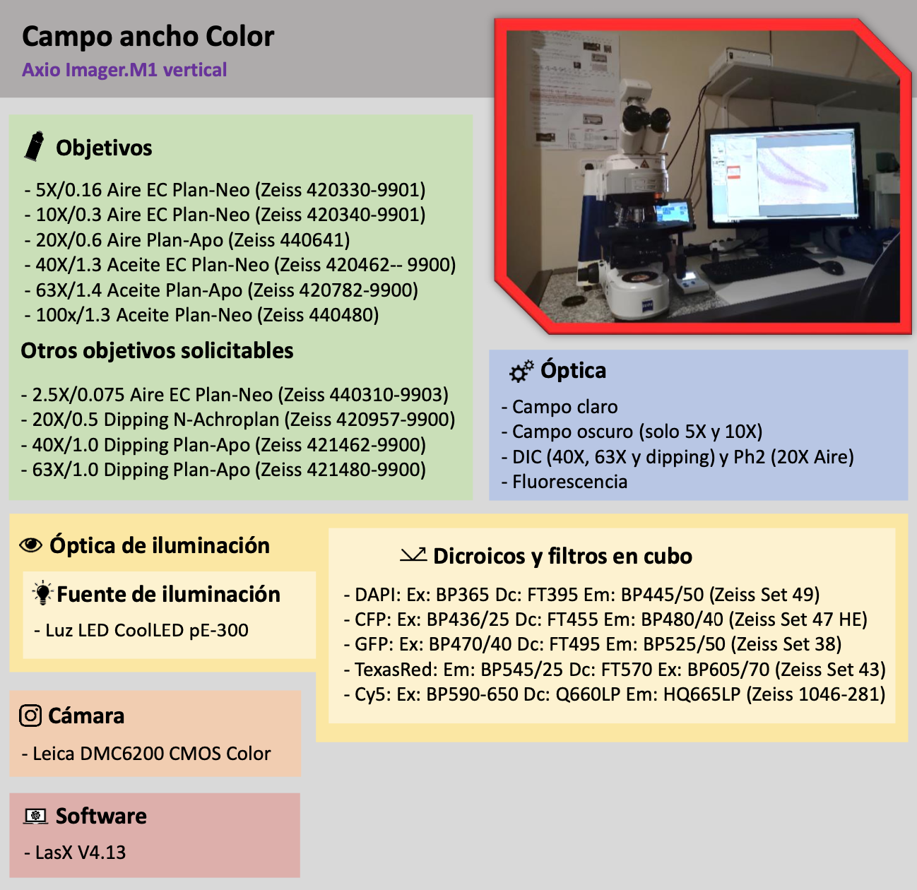
| Color Camera This simple upright system has a high-performance DMC6200 CMOS Color camera for color imaging, making it perfect for samples fixed with colorimetric stains such as hematoxylin/eosin, Xgal, Masson’s trichrome stain, etc. Although focused on color imaging, the system also has an LED light source and filters for fluorescence acquisition, so that a combination of both techniques is possible. Although focused on color imaging, the system also has an LED light source and filters for fluorescence acquisition, making it possible to combine both techniques. In addition, the system allows darkfield acquisition for those applications that require a better contrast than that generated by transmitted light in brightfield. |
 |
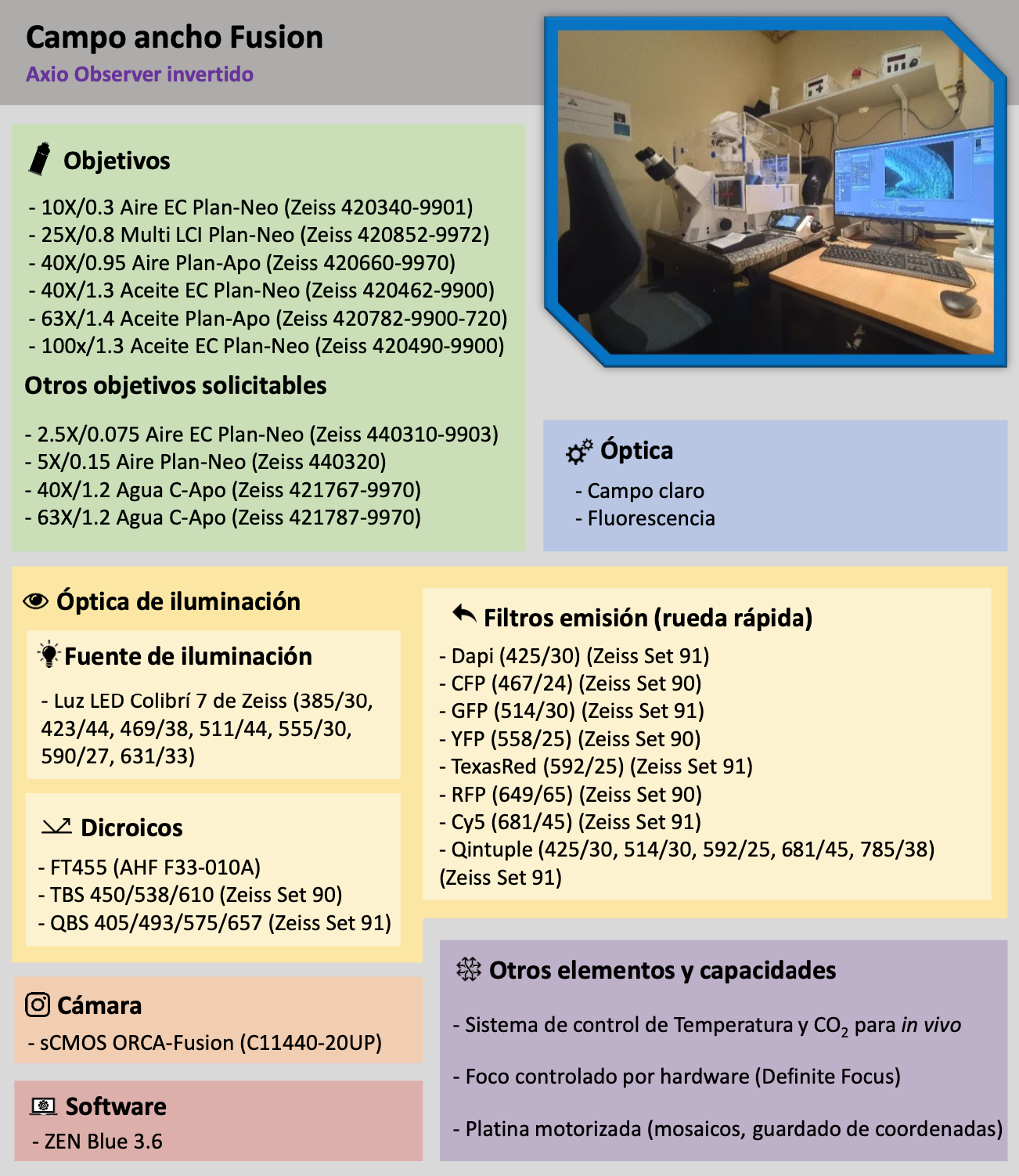
| Orca-Fusion Camera This equipment is our latest and most advanced widefield acquisition system. It has a state-of-the-art sCMOS camera (ORCA-Fusion) and a Zeiss LED light source capable of stimulating up to 7 ranges of the visible spectrum (Colibri 7). Its configuration of filters and dichroics allows a very high speed acquisition (up to 10-15 fps), suitable for very demanding experiments in terms of temporal resolution such as the study of calcium signals or the study of intracellular vesicles. In addition, it has the ability to control both the environmental conditions and the focus, making it perfect for long-duration in vivo experiment acquisitions. |
 |
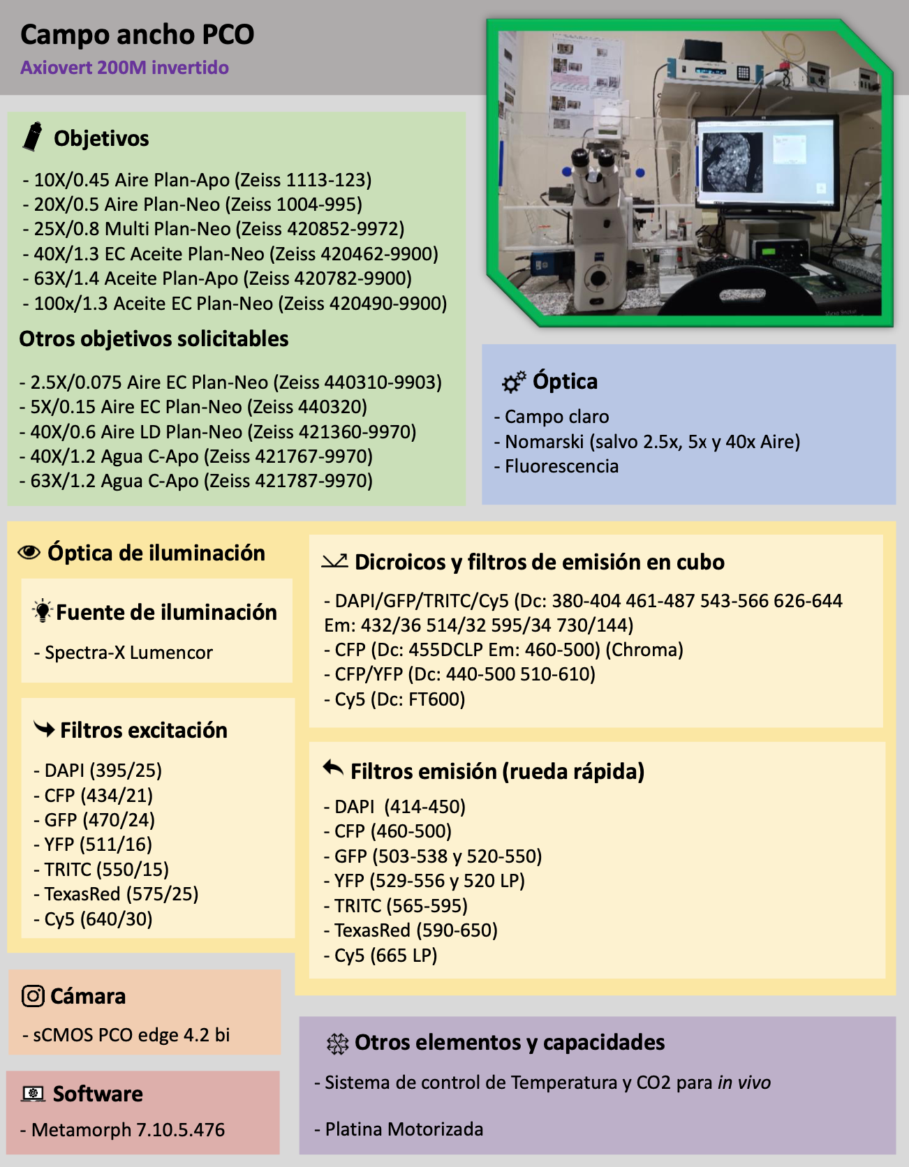
| PCO camera This wide-field system is controlled by the latest version of Metamorph software, has a highly sensitive sCMOS camera and the ability to control environmental conditions. Its LED light source and the versatility of its fast filter wheels with up to 10 positions make this system perfect for FRET (Förster resonance energy transfer) or for the acquisition of a wide range of fluorophores. |
 |
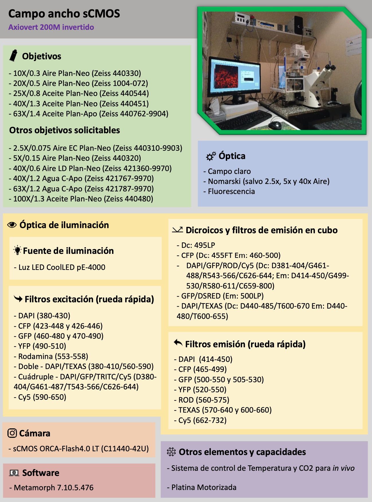
| Flash camera This system is similar in performance to the wide-field PCO system, with a highly sensitive sCMOS camera, the ability to control environmental conditions, a state-of-the-art LED light source with up to 16 excitation ranges and a high versatility in combining filters. It is therefore a fast, sensitive, robust system capable of supporting a high variability of fluorophores or specific experimental techniques such as FRET (Förster resonance energy transfer). |
 |
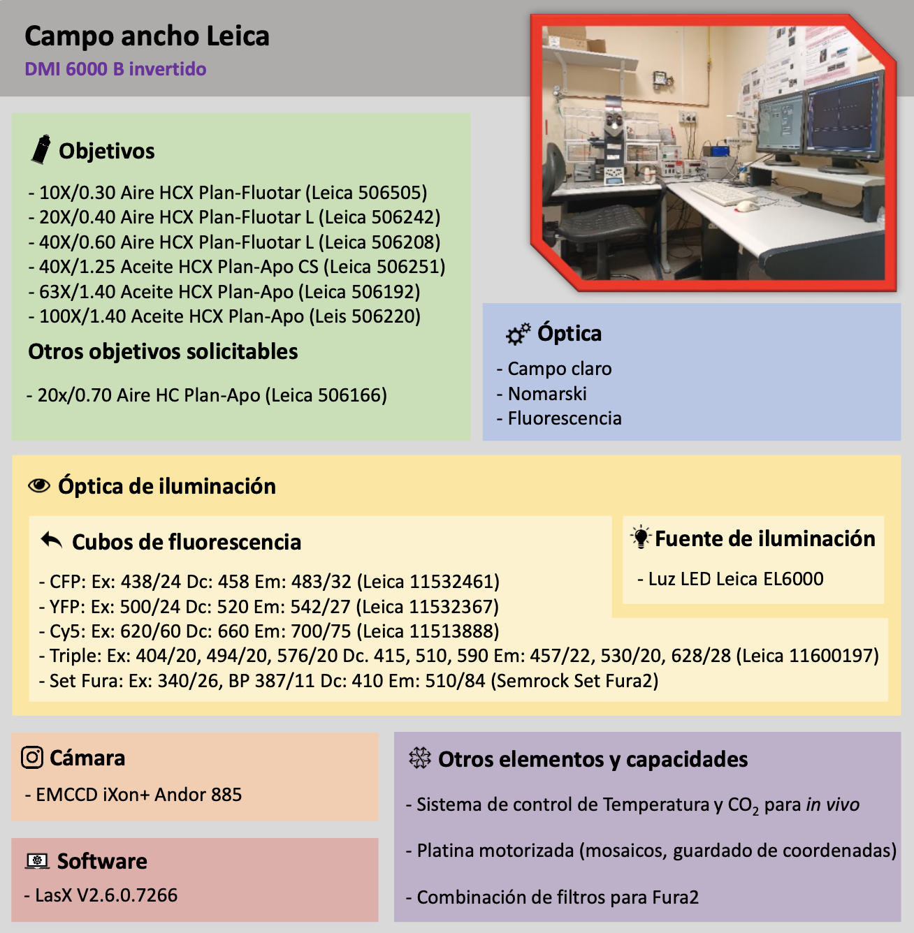
| Leica camera The Leica EMCCD widefield camera system is a simple-to-use instrument reserved for applications requiring low light conditions, very short exposure times and the highest sensitivity. The possibility to control the environmental conditions makes it perfect for in vivo experiments on very sensitive biological samples. In addition, it is the only system with filters suitable for calcium measurements through the Fura2 system. |
 |
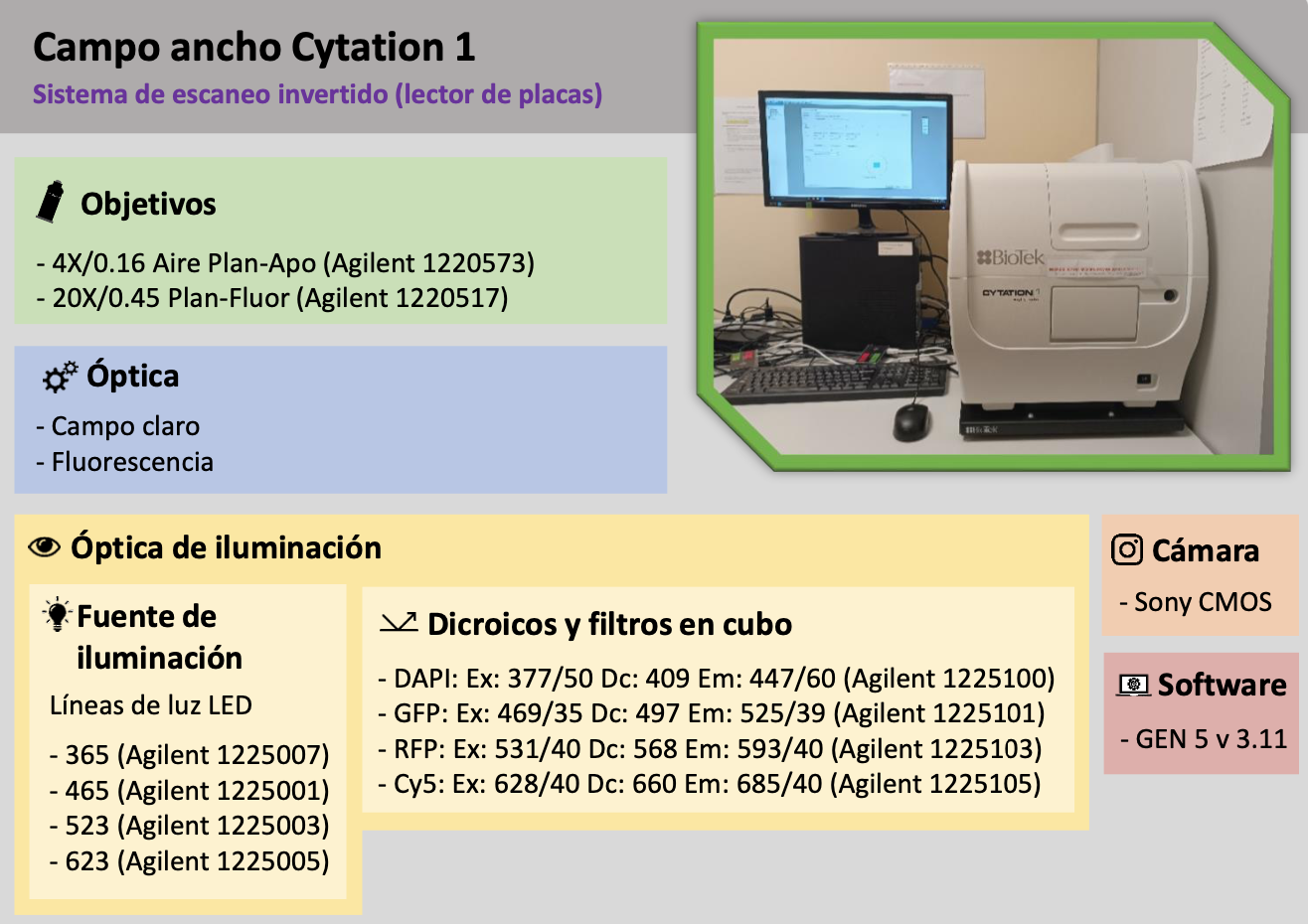
| Cytation 1 The Cytation1 is a simple widefield system with HCS (High Content Screening) capabilities for automated acquisition of multiwell plates. With two low magnification objectives (4x and 20x) and simple filters for the 4 most commonly used wavelengths (e.g. DAPI, A488, A555 and A647) it allows you to enter the plates and generate a protocol that quickly and automatically scans and acquires the desired wells in brightfield or fluorescence. The software also allows rapid subpopulation analysis for fast and automatic data acquisition. |
 |
Confocal Laser Scanning Microscopy
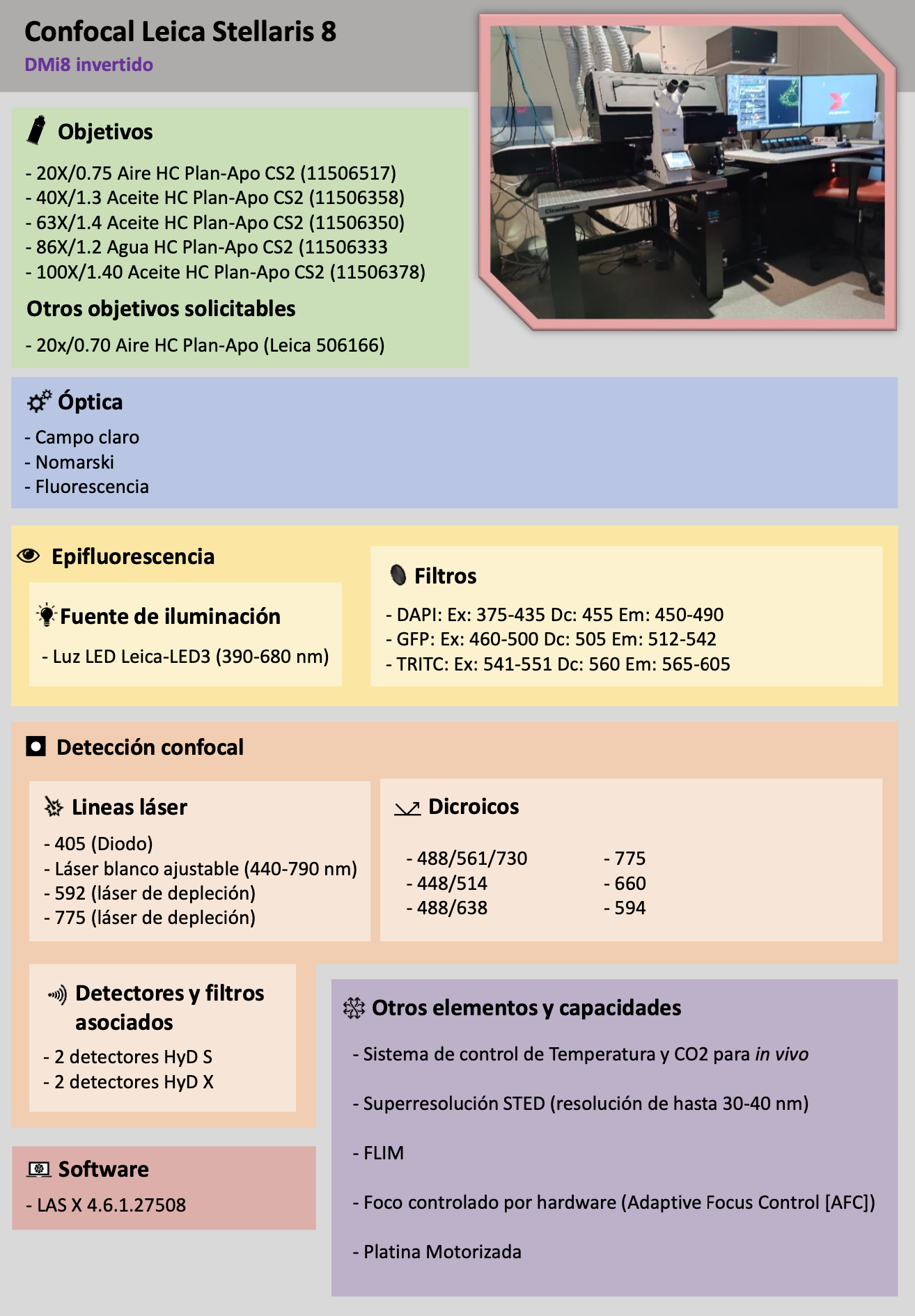
| Leica Stellaris 8 The Stellaris 8 system is one of Leica’s latest confocal models, easy to operate and with high image quality. This equipment features a white laser source that allows the use of the desired wavelength to maximize sample excitation, as well as the ability to perform in vivo experiments. The Leica Stellaris 8 is also equipped with a module for FLIM (Fluorescence Lifetime Imaging Microscopy) and two depletion lasers (592 nm and 775 nm) that allow you to acquire super-resolution images (30-40 nm) using STED (Stimulated Emission Depletion). |
 |
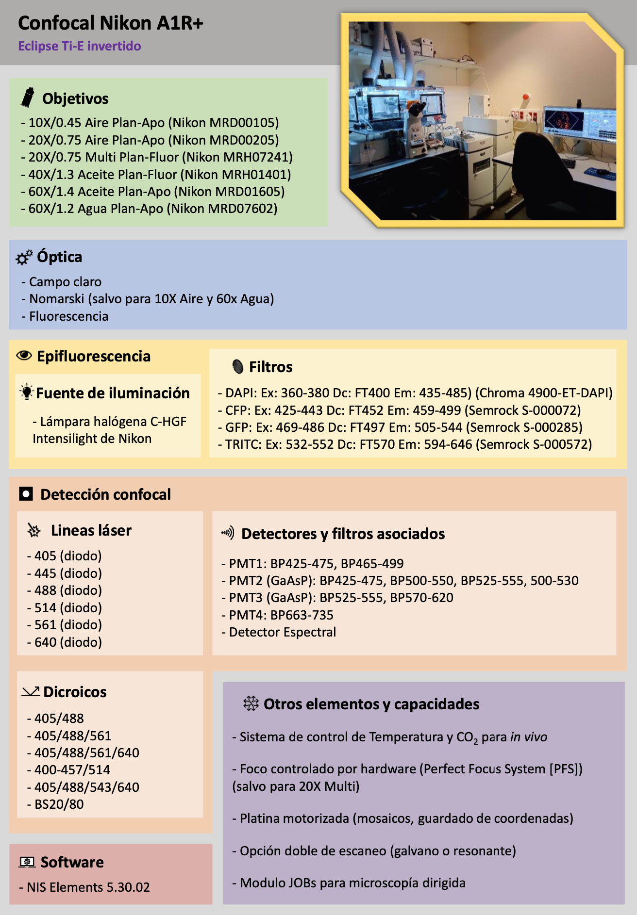
| Nikon A1R+ Nikon’s A1R+ is a reliable system with highly sensitive GaAsP detectors and six diode laser lines. This variety of laser lines and the presence of a spectral detector enable wide flexibility in fluorophore selection, allowing spectral acquisitions with signal spacings as small as 6 nm. This system also has a resonant scanner and the ability to control environmental conditions, making it perfect for in vivo and/or high-speed acquisitions without loss of confocality. The JOBs software module allows to automate acquisitions minimizing the need for user intervention. |
 |
|
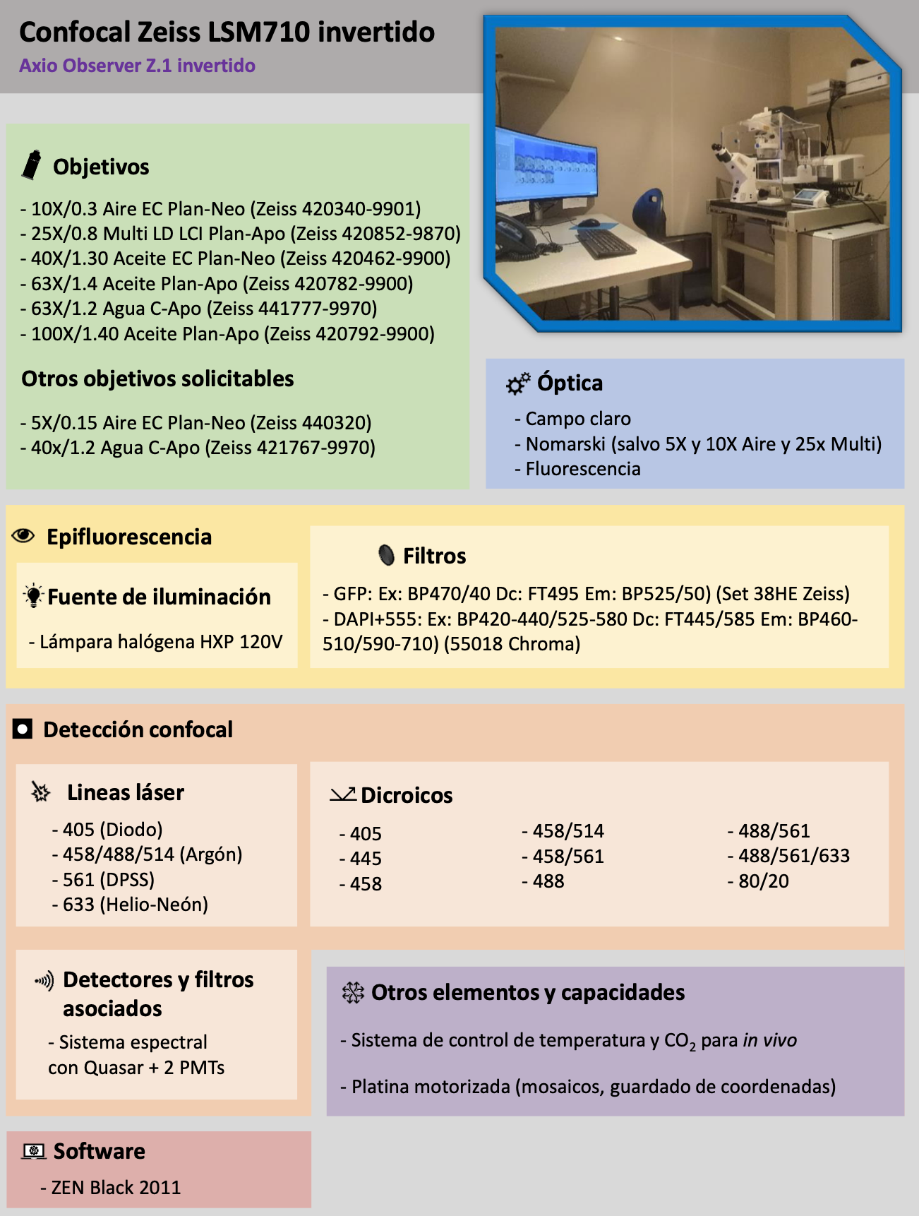
Zeiss LSM710 Inverted |
 |
| Zeiss LSM710 Vertical Like its inverted counterpart, the Zeiss LSM710 vertical confocal system has two spectral detectors and a total of six laser lines. It is therefore suitable for fixed samples that require flexibility in terms of the fluorophore panel. Its vertical stand also makes it perfect for dipping experiments, where the objectives have to be immersed directly into the sample medium itself, avoiding additional optical elements in the light path. |
 |
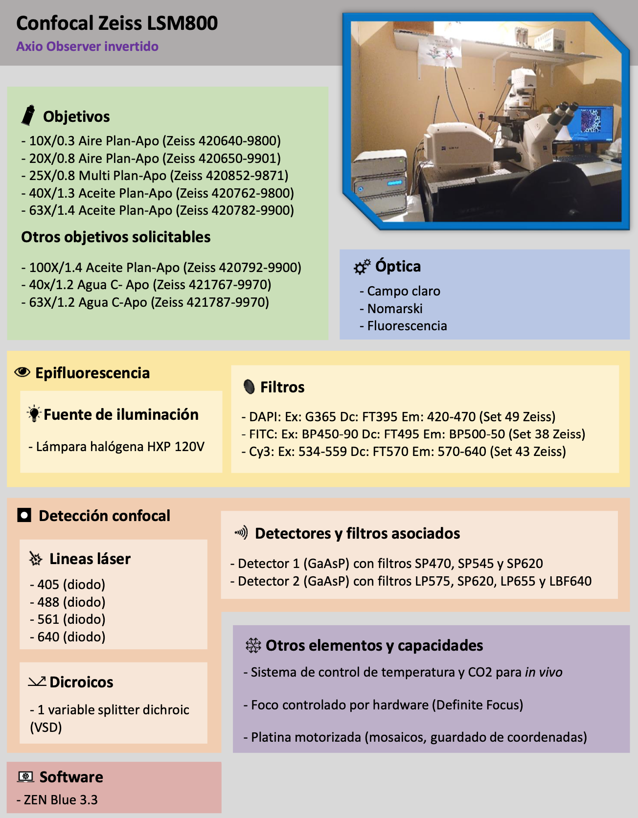
| Zeiss LSM800 The Zeiss LSM800 confocal microscope is a high-performance inverted system capable of controlling environmental conditions for in vivo experiments. It features high-sensitivity detectors and focus-holding capability for long-term acquisitions. |
 |
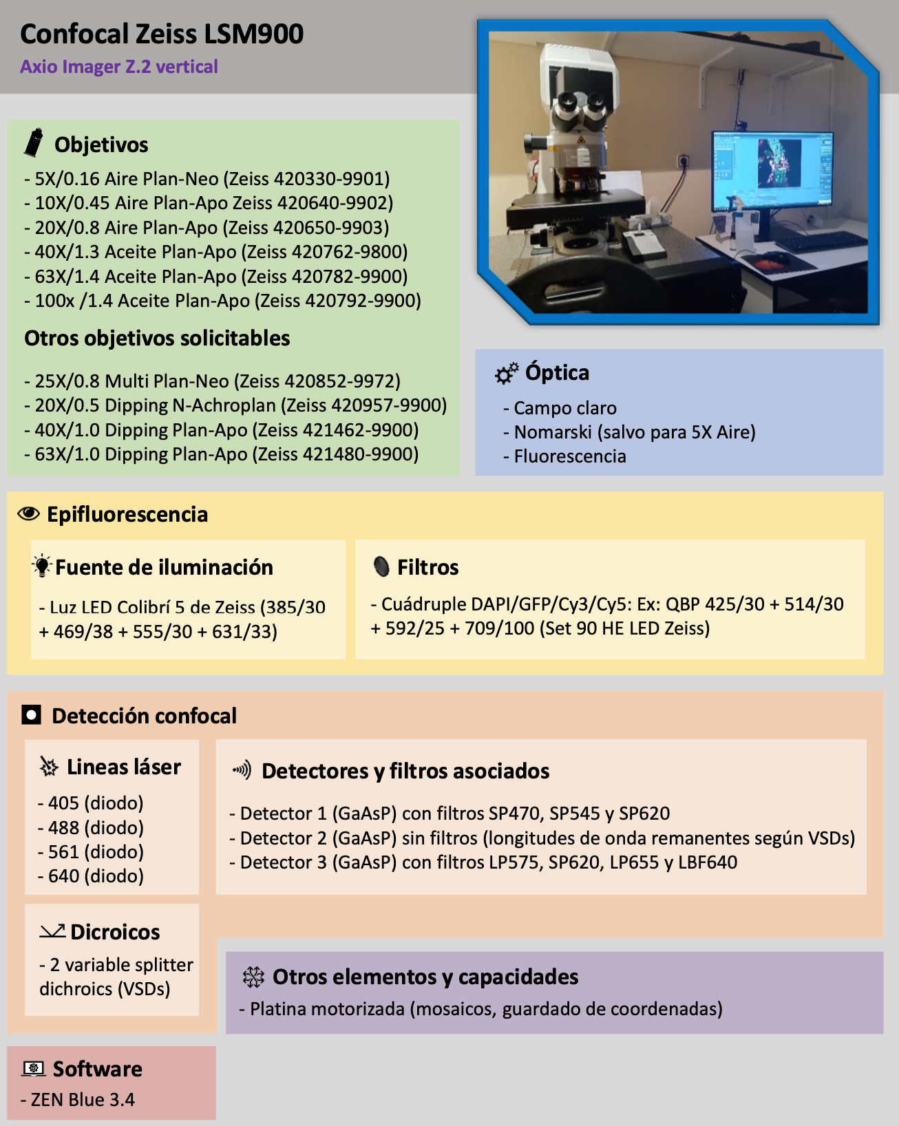
| Zeiss LSM900 The LSM900 confocal system is one of the latest generation systems from Zeiss. With high sensitivity detectors, this system has excellent optical quality and, being vertical, is a perfect system for fixed samples or dipping experiments requiring high image quality. |
 |
Confocal Spinning Disk Microscopy
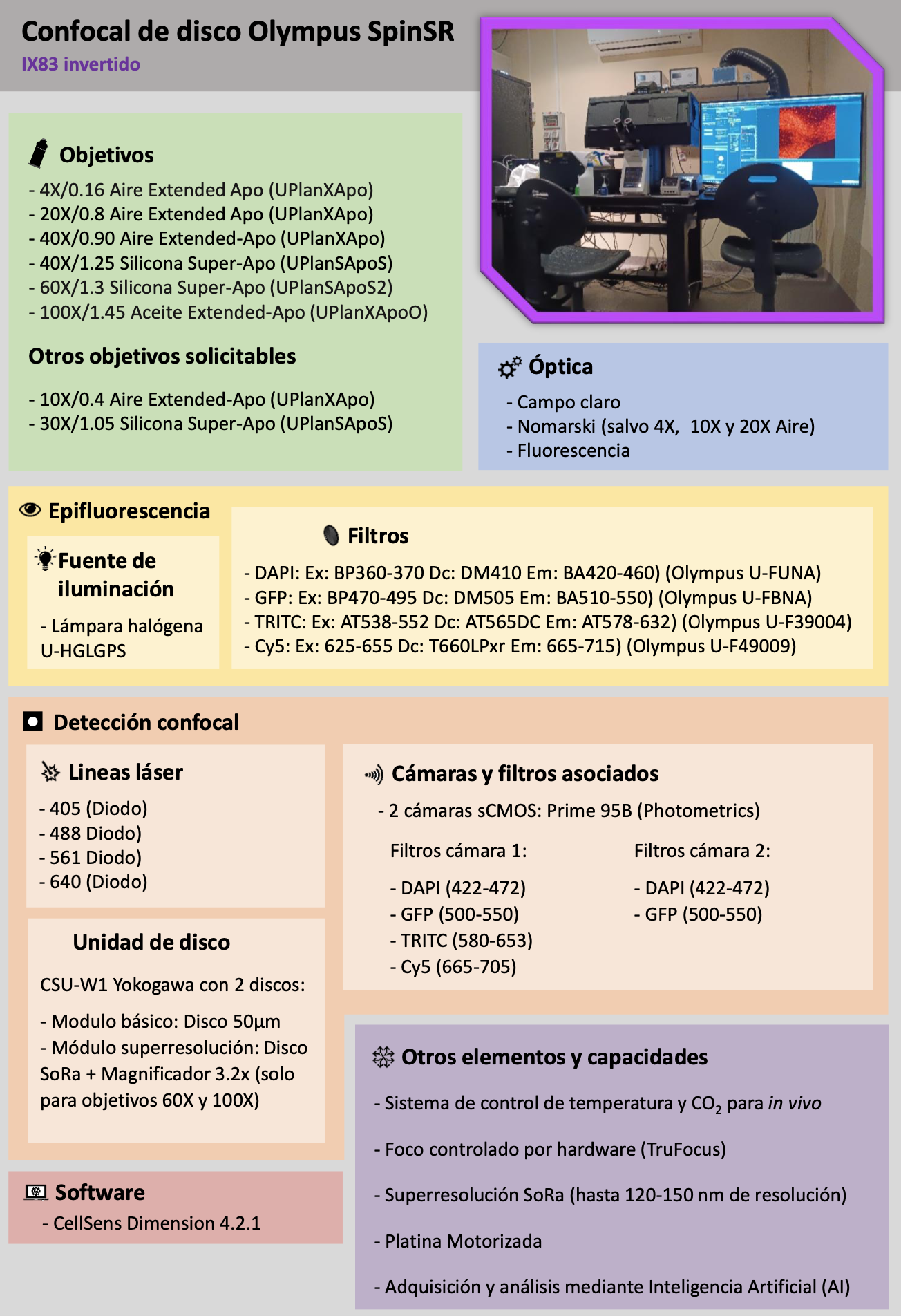
| Olympus SpinSR The Olympus SpinSR disk confocal system is one of our most sought-after systems, with four diode laser lines, two high-sensitivity sCMOS cameras for simultaneous two-color acquisition and the ability to control environmental conditions. It features high image quality at high speed while maintaining optical sectioning capability thanks to the latest version of the Yokowaga spinning disk unit (CSU-W1). In addition to a state-of-the-art spinning disk, this unit has an additional modified disk capable of acquisitions up to 120 nm resolution by optical photon remapping (SoRa). This unit also has a laser unit for FRAP (Fluorescence recovery after photobleaching) experiments at high speeds. |
 |
Lightsheet Microscopy
| ZEISS Z7 The ZEISS LightSheet Z7 system stands out for its outstanding performance in both fixed and cleared samples (using protocols such as iDISCO or CUBIC, among others) and in vivo applications, including zebrafish development, organoid research, and plant biology. The system features five laser lines (405, 445, 488, 561, and 647 nm), environmental control, two sCMOS cameras that enable simultaneous acquisitions, and a wide range of filters. These capabilities allow high-speed three-dimensional imaging of complex samples, whether live or fixed, with minimal phototoxicity. |
 |
Other optical equipment
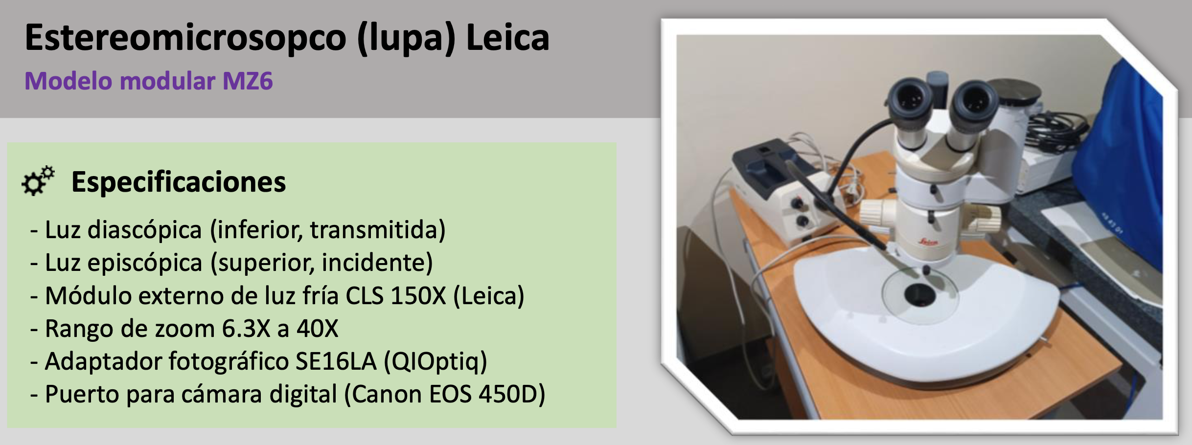
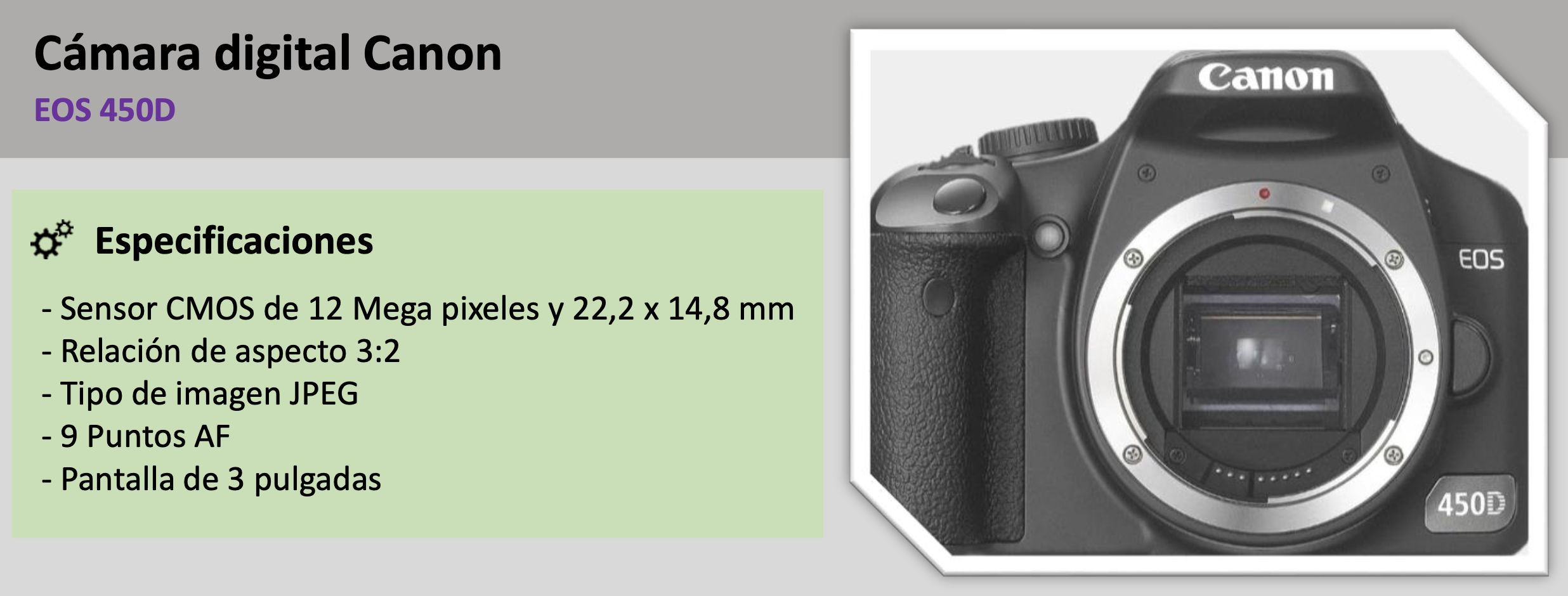
| Stereomicroscope (magnifier) Simple optical equipment with a maximum zoom of 40X and with external upper episcopic light (Leica CLS 150X cold light module) and lower diascopic light. Particularly suitable for the correct mounting of small samples or for general image acquisition thanks to its adapter for digital camera (Cannon EOS 450D). |
|
Analysis workstations
The service has two stations for the visualization and analysis of the acquired optical microscopy images. Both stations have 64 Gb of RAM memory, state-of-the-art graphics card and an 8-core processor. They also have 30-inch HP monitors.
 |
 |
Other equipment
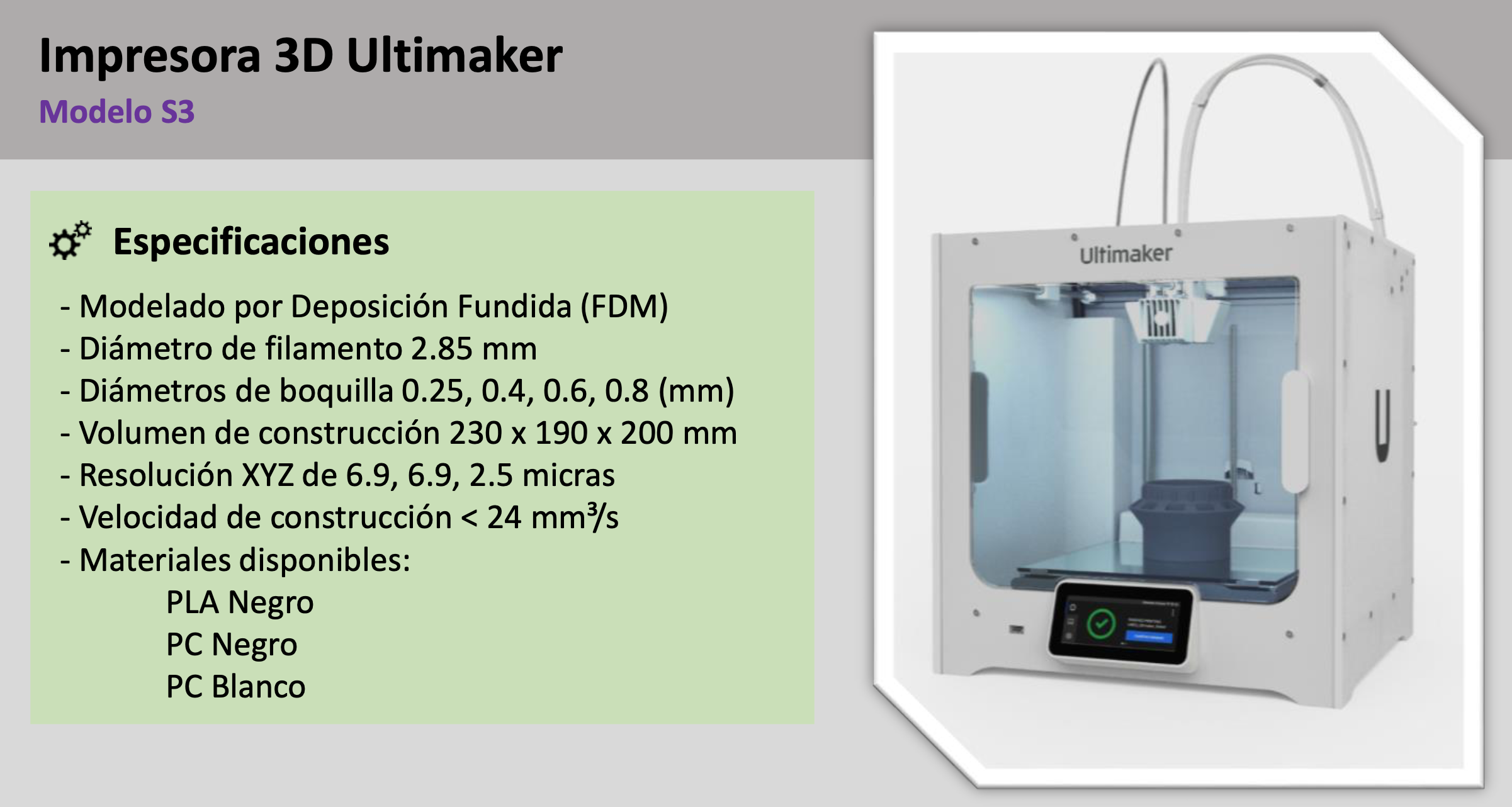
| 3D printer The unit has an Ultimaker S3 3D printer, capable of printing with a layer resolution of 20 microns and a total volume of up to 230 x 190 x 200 mm. Available materials to choose from are PLA Black, PC Black or PC White. |
 |
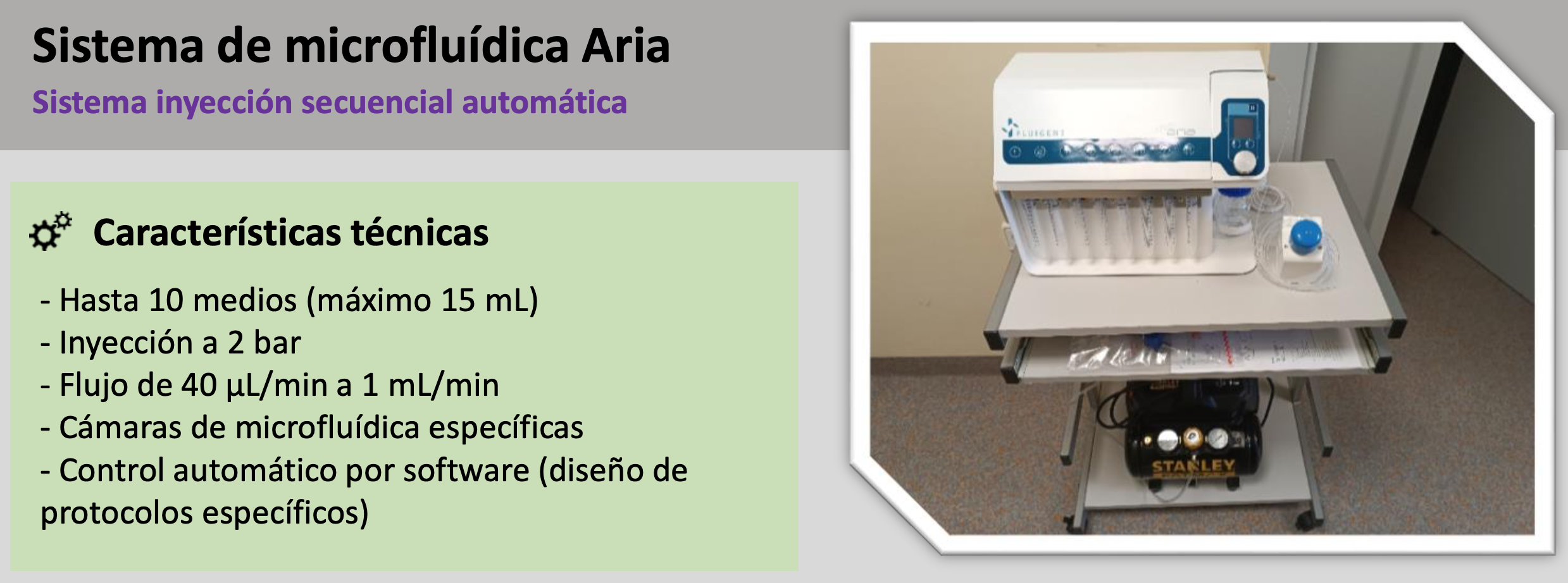
| Aria microfluidic system Fluigent’s Aria equipment is an automated sequential injection system very useful in experiments that require specific perfusion or injection of different media or components at specific times. This system allows to automate the addition of up to 10 different solutions into a microfluidic chamber or chip following previously defined protocols. |
 |
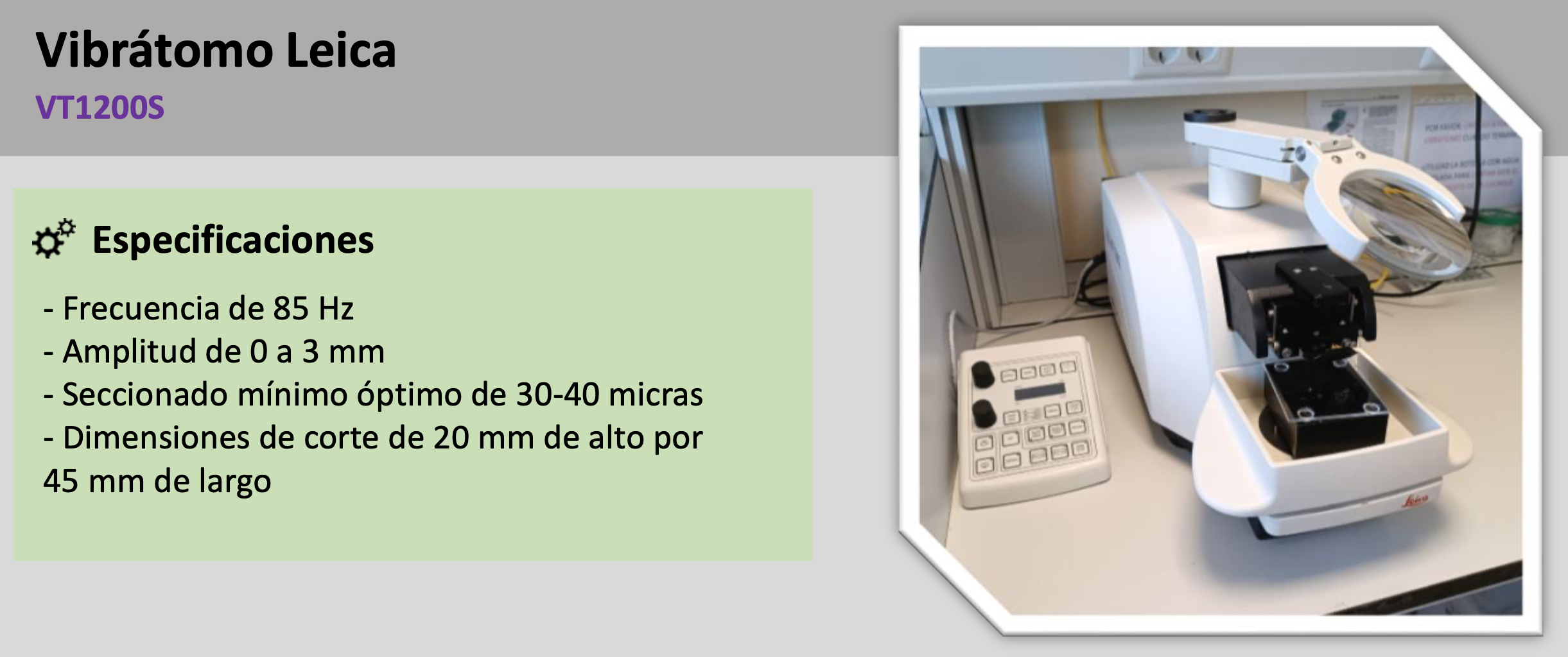
| Vibratome This equipment allows to section wet and soft samples (e.g. fresh tissues embedded in agarose) in a precise and automatic way. The sample size can be up to 30 x 40 mm and the section thickness can be adjusted in optimal increments with a minimum of 20-30 microns. It also has a magnetic sample holder, with which it is possible to change the sample orientation, and an integrated magnifying glass, which provides a better view of the cutting area. It also has an ergonomic and independent control panel. |
 |
Staff

María Teresa Villalba Villacorta
Lab.: 310 Ext.: 4643
tvillalba(at)cbm.csic.es

Carmen Sánchez Jiménez
Lab.: 310 Ext.: 4643
csjimenez(at)cbm.csic.es
Carlos Gallego García
Lab.: 310 Ext.: 4643
cgallego(at)cbm.csic.es

Elena Calvo Cazalilla
Lab.: 310 Ext.: 4643
elena.calvo(at)cbm.csic.es
Álvaro Sahun Español
Lab.: 310 Ext.: 4613
alvaro.sahun(at)cbm.csic.es
