Scientific facilities
Electron microscopy

The Electron microscopy facility provides scientific and technical support to research groups, both internal and external to the CBM, interested in using transmission electron microscopy for ultrastructural and immunocytochemical analysis of biological samples.
We can study macromolecular complexes, viruses, bacteria, eukaryotic cells and tissues. Available methods include conventional room temperature processing techniques and cryo-techniques such as cryosubstitution, cryoultramicrotomy and cryofracture.
Don´t even think of editing this!
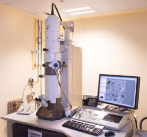
– Transmission electron microscope JEM1010 (Jeol,1992)
– Thermionic emission gun: tungsten filament.
– Accelerating voltage up to 100kV.
– Digital camera under CMOS column TemCam F416 (TVIPS)
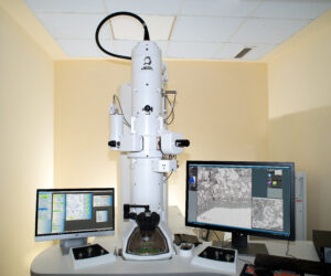
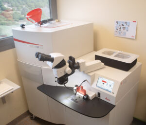
– JEM1400 Flash transmission electron microscope (Jeol, 2017)
– Thermionic emission gun: LaB6 filament.
– Accelerating voltage up to 120kV.
– OneView fast readout CMOS digital camera under column (Gatan).
Transmission electron microscopes
Transmission electron microscope JEM1010 (Jeol,1992)
– Thermionic emission gun: tungsten filament.
– Accelerating voltage up to 100kV.
– Digital camera under CMOS column TemCam F416 (TVIPS)
JEM1400 Flash transmission electron microscope (Jeol, 2017) Fot2
– Thermionic emission gun: LaB6 filament.
– Accelerating voltage up to 120kV.
– OneView fast readout CMOS digital camera under column (Gatan).
Cold sample processing equipment
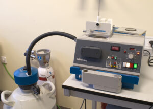
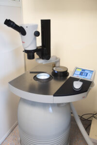
Cryofixation by immersion in cryogenic liquid (“plunge freezing” ), KF80 equipment (Reichert-Jung, Leica, 1992)
– Vitrification of particulate samples (macromolecules, viruses…) adsorbed on grids in liquid ethane.
– Cryofixation of chemically fixed and cryoprotected cells or tissues for processing by cryosubstitution or cryofracturing.
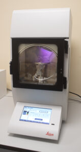
High pressure cryofixation (HPF), LEICA EM ICE (2023).
– Vitrification of cells, tissues and small organisms under native conditions for processing by cryosubstitution or cryofracturing.
Automatic cryosubstitution, LEICA EM AFS2 (2023).
– Dehydration at low temperatures, -90 °C, -80 °C, cryoinclusion in Lowicryl resins and polymerization with UV light at low temperatures.

Cryofracture/ Freeze-etching, BAF060 equipment (Leica, 2007)
– Cryofracture of cells and tissues and obtaining metallic replicas of the fractured surface.
– Pt/C rotary shading of nucleic acids and viruses.
Ultramicrotomes
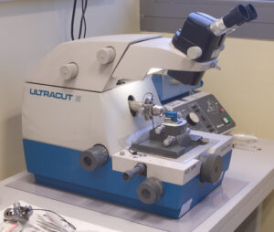
Ultramicrotome ULTRACUT E (Reichert-Jung, Leica)
– to obtain semithin, 1µm, and ultrathin, 50nm – 90nm sections at room temperature of samples embedded in resins.
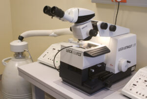
Ultramicrotome with cryo camera, UCT (Leica)
– For semithin, 1µm, and ultrathin, 50nm – 90nm sections at room temperature of samples included in resins.
– To obtain cryosections at low temperature, – 80 ºC to -120ºC for immunolabeling on thawed sections (Tokuyasu technique).
Leica EM AC600 Evaporator (2023)
– Evaporation of a carbon film with defined thickness on grids.
– Ionization of grids for negative staining with “Glow discharge”.
Techniques
– Negative staining of macromolecular complexes and viruses. Photo technique 1
– Analysis of nanoparticles.
– Conventional embedding of biological samples in resins and ultramicrotomy. Technical photo 2
– Cryofixation by immersion in liquid ethane and high pressure.
– Cryosubstitution and inclusion at low temperature. Technical photo 3
– Electron immunomicroscopy with colloidal gold conjugates in resin sections. Technical photo 4
– Electron immunomicroscopy with colloidal gold conjugates in cryosections (Tokuyasu technique). Technical photo 5
– Cryofracture, cryosectioning and obtaining metal replicas. Technical photo 6
– Electron microscopy of nucleic acids. Technical photo 7
– Correlative optical-electron microscopy (CLEM).
Staff
Milagros Guerra Rodríguez
Lab.: 113 Ext.: 4532
mguerra(at)cbm.csic.es
Tania Matamoros Grande
Lab.: 113 Ext.: 4532
tmatamoros(at)cbm.csic.es
Raquel Losada de Paz
Lab.: 113 Ext.: 4538
rlosada(at)cbm.csic.es

Martin Christoph Sachse
Lab.: 113 Ext.: 4538/4532
martin.sachse(at)cbm.csic.es